
Hydroxyapatite is a mineral that is found in bones. Its crystalline structure is similar to that of human bone, and it helps promote new bone formation. It can also be used to coat implants, making them more biocompatible.
Adding HA to polymers makes it possible to develop a biodegradable scaffold for bone tissue engineering. Ion doping of HA improves its biological properties.
Osteogenesis
Hydroxyapatite can induce bone formation at the cellular and organic levels. It is often deposited on titanium oxide (TiO2) surfaces to promote osseointegration of biomedical implants. This process is called “biomimetic” because it mimics the natural remineralization of bones. During this process, hydroxyapatite deposits onto the surface of TiO2, enhancing its bonding to bone. It also increases the speed at which bone ingrowths are formed. Its low crystallinity and porous structure make it an ideal candidate for this role.
The skeletal components of the vertebrae, pelvis, and limbs are initially formed from cartilage tissue that serves as a model for the bone that will eventually form. During the intramembranous ossification process, the cartilage is degraded and osteoblasts modify the cartilage matrix to include an osteoid scaffold. The hydroxyapatite in the osteoid staging acts as a mineral matrix that helps to form new bone tissue. The osteoblasts are surrounded by stromal cells that can secrete calcifying substances such as collagen and osteocalcin.
Osteogenesis imperfecta (OI) is a genetic condition that causes fragile, easily broken bones. The disorder is characterized by a defect in the gene that makes collagen, the protein that holds the mineral parts of bone together. It is also known as brittle bone disease and can cause severe problems.
People with OI experience frequent fractures, even after mild injury or without a clear cause. In most cases, OI affects the ribs and hip bones. It can also cause problems with the muscles, joints, eyes, ears, skin, and blood vessels. In addition, some types of OI can affect the teeth (dentinogenesis imperfecta).
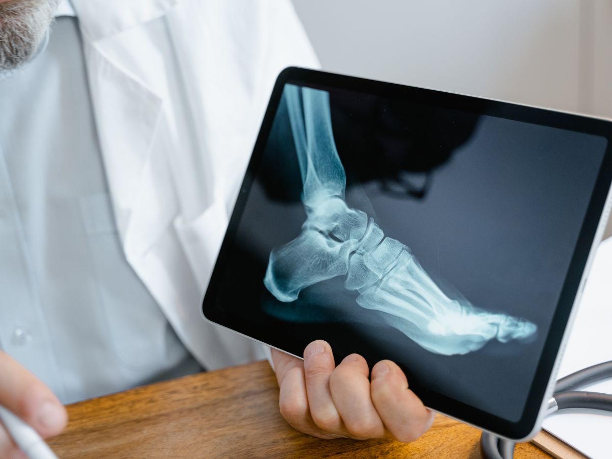
The simplest way to diagnose OI is to do DNA testing. This test will detect the defective genes that are responsible for OI. The mutations usually occur in the COL1A1 or COL1A2 gene. This testing is very accurate and can be done quickly and easily. Other tests, such as X-rays and bone scans, can help determine the OI’s severity. These tests can detect the onset of OI and permit to identify patients who may require surgery or other treatments.
Osseointegration
Hydroxyapatite is a natural mineral that exists in living bone. Its natural form consists of a complex structure that contains a calcium-phosphate crystal system. It has the formula Ca10(PO4)6(OH)2. It is a polymorphic compound with several variations of the structure. These variations occur because of substitutions of the ions in the crystal system. For example, if calcium is replaced by strontium, the compound becomes Ca10Sr(PO4)6(OH)2. This compound has many uses in medicine and dentistry.
It is used in bone substitutes and scaffolds to repair fractured bones. It can also be used to create a surface on orthopedic and dental implants to promote growth of bone tissue. HA can be obtained from biological (coral, bovine or marine algae-derived) sources or synthetically. It is available as granules and blocks, by itself or as a composite with polymers or other ceramics, or as a coating on orthopedic or dental implants.
The hydroxyl ions in hydroxyapatite are bonded to the crystal structures and act as an anchor point for other chemical compounds in bone. This makes HA a strong, durable material. Its porosity increases its bioactivity, especially in the presence of blood vessels. The pore size of the apatite influences protein adsorption and the ability to absorb calcium and phosphate. The porosity of HA also impacts its mechanical strength.
Osseointegration is a surgical technique that allows limb amputees to attach their prostheses directly to the bone of the residual limb, eliminating socket-related problems like pain, discomfort and swelling. It has revolutionized the lives of thousands of patients around the world.
This unique process was first developed in the 1950s by Swedish physician Dr. Per-Ingvar Branemark. Since then, it has become an essential part of modern orthopaedics. Its success has transformed patients’ lives with lower and upper limb amputations. Osseointegration enables these patients to live more active lifestyles and regain the feeling of full participation in activities. It is possible to learn more about this life-changing procedure by visiting a clinic that offers it. A team of specialists in orthopaedics, plastic surgery and prosthetics performs osseointegration surgery.
Bone mineral density
Hydroxyapatite is an important inorganic material found naturally in bone, tooth enamel and dentine. It has the chemical formula Ca10(PO4)6(OH)2. The crystal structure of hydroxyapatite is a tetrahedral tetrahedron. Strong, ionic hydrogen bonds bind together its phosphate and calcium atoms.
The main function of hydroxyapatite in bone is to provide a matrix for mineral deposition. Hydroxyapatite promotes osteogenesis during bone formation by providing the structural framework necessary for mineral deposition and stimulating cell adhesion. In addition, hydroxyapatite contains many ionic sites, which allows it to release calcium and phosphate in response to stimuli such as cell signaling.
Visit our product hydroxyapatite bone graft: Phase Pure Hydroxyapatite
In humans, hydroxyapatite is primarily present in the bones of the lower jaw and the hips. The bones of the arms and legs contain hydroxyapatite in smaller amounts. Hydroxyapatite provides a hard, dense, porous scaffolding that fuses with the adjacent bone to form dense bone during growth and development. Hydroxyapatite is also deposited in the joints of the upper and lower extremities to support ligaments and tendons.
Bone mineral density (BMD) is an important measurement of bone strength. A bone density scanner, or a special x-ray can measure BMD. Generally, a higher BMD is associated with a lower fracture risk. However, this is not always the case. In some populations, fractures reach very high levels despite low BMD. The rate of bone loss is also more significant at certain times in the life cycle, such as during menopause, when levels of the bone-bolstering estrogen fall.
Hydroxyapatite has been used extensively in orthopedics as a biomaterial to promote tissue regeneration. It is especially attractive because of its low cost, abrasion resistance and osteoinductive properties. In the laboratory, it has been shown to induce bone regeneration with minimal vascular invasion.
In some cases, calcific hydroxyapatite deposits may be mistaken for tumors or infections. Because of this, radionuclide scanning is often performed to establish the diagnosis. In some cases, the use of a radiotracer such as technetium-99m helps distinguish HADD from other calcific processes by measuring the uptake of the tracer in the area of the deposit.
Bone regeneration
Bone regeneration is a complex biological process when a bone fractures. It includes three intersecting phases: inflammation, vascularisation, and bone production. This process requires many progenitor cells, including osteoblasts, endothelial, and hematopoietic stem cells. The primary function of these cells is to form the matrix and provide a structural foundation for new bone formation. In addition, they also participate in other vital processes, such as the transport of oxygen and nutrients to the fractured area.
Hydroxyapatite (HA) is one of the main inorganic components of normal human bone. It is bounded to the organic matrix by a network of protein molecules. This enables the integration of the HA crystals into the bone, which provides a strong mechanical and biochemical support. The hydroxyl groups on the surface of HA are reactive and can bond with protein molecules. This is why HA is an excellent candidate for bone substitutes.
In vitro studies have shown that porous HA can accelerate bone ingrowths onto titanium implants. This is because it provides a biomimetic environment that mimics the natural physiology of the human body. This is important because it may improve the bone-bonding ability of titanium. In addition, it can enhance the biomechanical properties of the implant.
A recent study has found that the amount of HA in the organic portion of the bone is directly related to its strength and hardness. This is believed because the HA crystals are arranged in a tetrahedral pattern. The tetrahedral arrangement of HA crystals also contributes to its high porosity, another factor contributing to its strength and durability.
Although the exact function of HA in the bone is still not fully understood, it is clear that it plays an essential role during bone healing and osseointegration. It is therefore vital that we continue to learn more about the biochemical processes that occur at a cellular and molecular level during bone regeneration and develop techniques for measuring these parameters in vivo. This will allow us to develop more effective bone replacement and renewal strategies.
Read More: THE ULTIMATE GUIDE: HOW TO USE PAIN O SOMA 500MG FOR MUSCLE PAIN RELIEF